Displasia Acetabular: La Causa Oculta de tu Dolor Crónico de Cadera
Acetabular Dysplasia: The Hidden Cause of Your Chronic Hip Pain
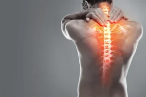
If you have chronic pain in your groin or hip, especially if you are young or middle-aged, it is likely not simply wear and tear from aging. Behind that persistent pain, which limits your life and prevents you from participating in sports, there may be a structural condition that many general physicians overlook: acetabular dysplasia.
What Is Acetabular Dysplasia and Why Does It Cause Pain?
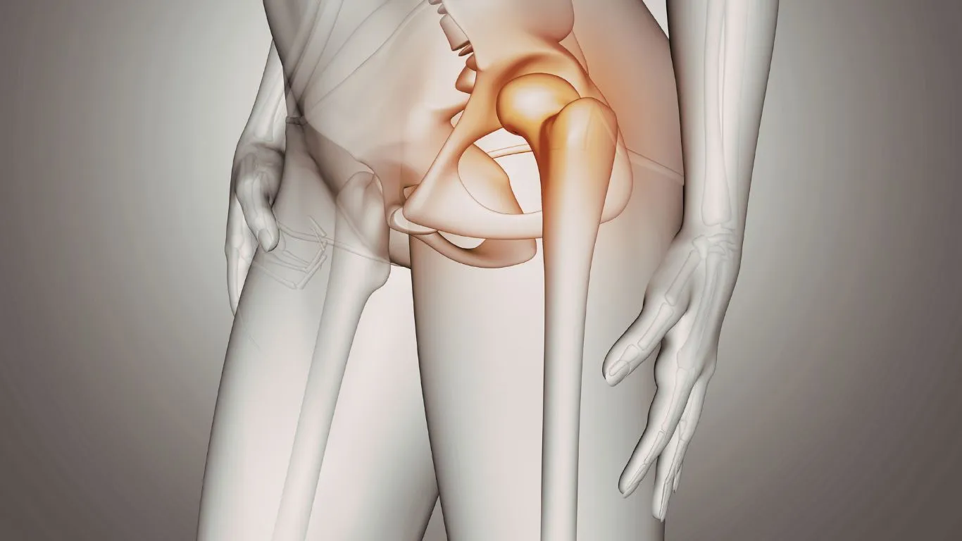
Acetabular dysplasia is a malformation of the hip that develops during growth. Imagine your hip joint as a ball (the femoral head) that fits into a socket (the acetabulum). In a normal hip, the acetabulum is deep and fully covers the ball, distributing weight evenly.
However, if you have dysplasia, your acetabulum is too shallow or incorrectly angled. In other words, the roof that should cover and protect the femoral head is insufficient. As a result, body weight is not distributed across the entire contact surface. Instead, it is concentrated in a very small area, usually at the upper edge of the acetabulum. This leads to constant overload.
This chronic overload is the driving force that accelerates cartilage wear. Consequently, the joint begins to deteriorate prematurely, initiating a painful process known as osteoarthritis or Hip OsteoarthritisIf we do not correct the structure, the cartilage will continue to suffer irreversible damage.
How Do I Know If I Have Acetabular Dysplasia? Key Symptoms
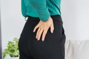
The pain associated with acetabular dysplasia is usually insidious, meaning it starts mildly and progressively worsens over time. Patients often report a sensation of instability or that the hip gives way under pressure. It is essential that you pay attention to the location and type of pain so that we can make a timely diagnosis.
Groin Pain and Anterior Thigh Pain
This is the most common symptom. The pain is located deep in the groin and may radiate to the front of the thigh or even down to the knee. Initially, the pain appears only during intense activities, such as running or practicing high-impact sports. However, as the condition progresses and cartilage damage increases, the pain becomes constant.
Functional Limitation and Stiffness
You will begin to notice that everyday activities become difficult. For example, climbing stairs, getting up from a low chair, or putting on your shoes may cause sharp discomfort. In addition, morning stiffness is common. At the beginning of the day, your hip feels stiff and it becomes difficult to start moving normally. This functional limitation is a clear sign that the joint is struggling to move freely.
Sensation of Instability or Clicking
Due to the lack of adequate bony coverage, the femoral head may move slightly more than normal. This can create a sensation of instability or cause the hip to pop or click painfully, especially during rotation or pivoting movements. This instability may indicate associated damage to the labrum, the sealing structure that surrounds the acetabulum.
Causes and Mechanics of Premature Wear
Acetabular dysplasia is classified as a primary cause of Hip Osteoarthritis in young adults. It is crucial to understand that it is not a condition acquired from poor habits, but rather a failure in bone development. The main cause is developmental dysplasia of the hip (DDH) that was not fully detected or corrected during childhood.
Abnormal Biomechanics
The central problem is focal overload. When the acetabulum is shallow, the joint is constantly under stress. The force that should be distributed over 10 cm² becomes concentrated in 2 cm². This is similar to walking in high heels instead of flat shoes; the pressure per square centimeter increases dramatically. As a result, the cartilage, which acts as the natural shock absorber, degenerates much faster than it should. This leads to early hip osteoarthritis, sometimes even before the age of 40.
General Risk Factors
Although the structural abnormality is the root cause, other factors can accelerate the degenerative process. For example, excess weight increases axial load on the dysplastic hip. In addition, high-impact sports or activities involving repetitive pivoting, such as soccer or ballet, can worsen symptoms.
However, correcting these factors is secondary; the primary focus must be on correcting the structural problem.
Accurate Diagnosis: The Key to Joint Preservation
As a specialist in joint preservation, my goal is to diagnose dysplasia before cartilage damage becomes irreversible. The diagnosis of acetabular dysplasia is based primarily on a thorough physical examination and highly specific imaging studies.
During your consultation, I will evaluate your range of motion, your gait, and look for signs of pain during specific maneuvers. However, confirmation is radiological.
We need a high-quality standing anteroposterior (AP) pelvic X-ray. In this image, we not only evaluate bone shape but also measure specific angles that determine the level of coverage. We assess the Wiberg angle (lateral center-edge angle) and the Tönnis angle. If the Wiberg angle is less than 20 degrees, coverage is insufficient and the diagnosis of dysplasia is confirmed.
In some cases, if we suspect cartilage or labral damage, or for precise surgical planning, we will use magnetic resonance imaging (MRI) or computed tomography (CT). MRI allows us to evaluate soft tissues and cartilage, which is essential to determine prognosis and the most appropriate treatment for you.
Treatment Options for Acetabular Dysplasia: From Observation to Joint Preservation Surgery
The treatment of acetabular dysplasia depends fundamentally on two factors: the patient’s age and, most importantly, the degree of arthritic damage (hip osteoarthritis) already present in the joint.
Conservative Treatment: Symptom Management
If dysplasia is mild and has not yet caused significant cartilage damage, we may attempt to manage symptoms. Conservative treatment includes:
- Specialized physical therapy: Strengthening the periarticular muscles, especially the hip abductors and flexors, can help stabilize the joint and partially compensate for the lack of bony coverage.
- Activity modification You must avoid high-impact sports and activities involving sudden turns or heavy loads. This reduces stress on the cartilage.
- Medications Nonsteroidal anti-inflammatory drugs can relieve pain, but it is crucial to understand that they only treat the symptom, not the structural cause of the disease. Injections with hyaluronic acid may provide temporary relief, but they do not stop the progression of osteoarthritis.
I must be clear: conservative treatment is a temporary measure. If the bone structure is incorrect, degeneration will continue. The only way to stop osteoarthritis progression is by correcting the anatomy.
Surgical Treatment: Periacetabular Osteotomy (PAO)
Periacetabular osteotomy is the gold standard and the procedure of choice to correct acetabular dysplasia in young, active patients who do not yet have severe osteoarthritis. This procedure represents the essence of joint preservation.
Periacetabular osteotomy is a complex and highly specialized surgery. It involves making controlled cuts around the acetabulum (the socket) to free it from the pelvis. Once released, the acetabulum is repositioned. It is rotated and moved laterally to better cover the femoral head. Finally, it is fixed in its new position with screws.
The goal of this procedure is to redistribute load across a larger and healthier joint surface. By doing this, we eliminate the overload point and stop—or significantly slow—the progression of osteoarthritis. When performed at the right time, periacetabular osteotomy can save your hip and delay the need for a total hip replacement for decades.
Total Hip Replacement
If dysplasia has been ignored for too long and osteoarthritis has already destroyed much of the cartilage (advanced osteoarthritis), periacetabular osteotomy is no longer a viable option. In these cases, the only effective solution to eliminate pain and restore function is total hip replacement. Although this is an excellent and durable solution, my priority as a joint preservation surgeon is to help you avoid it whenever possible through early structural intervention.
If you identify with these symptoms, or if you have already been diagnosed with dysplasia, do not delay your decision-making. Every month that passes without correcting the biomechanics is a month in which your cartilage continues to wear down. Acting in time is the difference between preserving your joint and needing a total replacement.
I invite you to schedule an evaluation with me. We will analyze your X-rays, measure your angles, and design a personalized treatment plan. Together, we will work so that you can regain your quality of life.

Dr. Raul Lopez Solis
Hip & Knee Surgeon
Professional License: 926463 / Health Ministry Registry (SSA - Mexico): 2204 / Specialty License No.: AESSA-27436
Facebook: Dr Raul López Orhtopedic Surgeon
Instagram: drraullopezs
TikTok: @drraullopezs
Web site: About me
Related posts